

Real Treatment.
Real Relief.
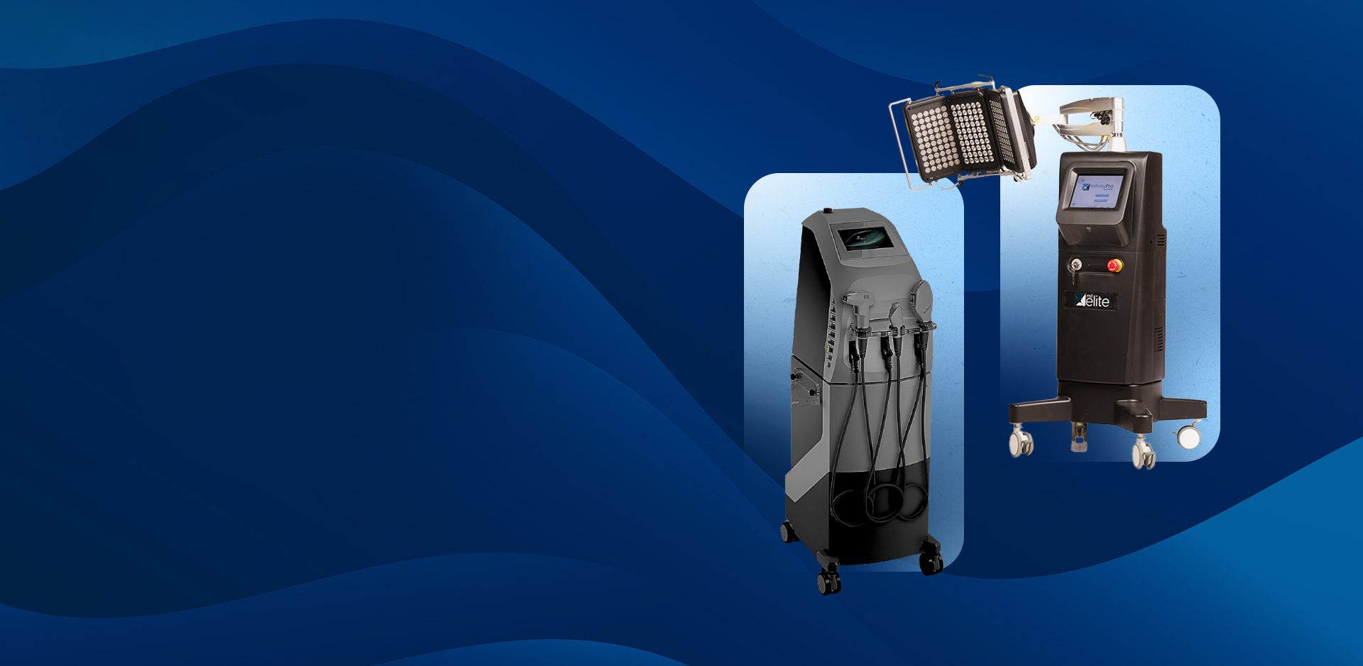
Dry eye disease has a root cause — and at Lighthouse Vision, we treat it. Using a comprehensive suite of advanced therapies, we improve meibomian gland function, reduce inflammation, and rebuild a healthy tear film that lasts.
9+ Treatment
Modalities
60 min Comprehensive
Evaluation
17+ Oculus 5M
Diagnostic Tests
IPL + RF Advanced
Energy Therapies
Insurance Accepted for
Medical Visits
The root cause
Most dry eye is caused by
blocked glands, not a lack of tears.
The most common form of dry eye is Meibomian Gland Dysfunction (MGD) — the tiny oil-producing glands along your eyelid margins become clogged, damaged, or inflamed. Without this protective oil layer, your tears evaporate too quickly and your eyes cannot maintain a stable, comfortable surface.
Dry eye is also driven by chronic inflammation, hormonal changes, systemic conditions like Sjogren’s syndrome and rosacea, and neurological factors. Understanding the specific mechanism in each patient is why we begin every treatment relationship with a comprehensive diagnostic evaluation — not a one-size-fits-all approach.
Common Symptoms
Burning, stinging, or gritty sensation
Light sensitivity (photophobia)
Blurred vision that fluctuates with blinking
Eye fatigue — especially with screens or reading
Redness or persistent irritation
Contact lens intolerance or dropout
Watery eyes (reflex tearing in response to dryness)
Difficulty in wind, low humidity, or air conditioning
MGD
is responsible for up to 86% of all dry eye disease
Your first visit
A Comprehensive Dry Eye Evaluation
Every patient begins with a dedicated 60-minute dry eye evaluation — one of the most thorough ocular surface assessments available in Connecticut.
We use objective, reproducible testing to identify exactly what type of dry eye you have and which structures are involved before recommending any treatment.
Powered by
Oculus 5M Keratograph
Our evaluation is anchored by the Oculus 5M — one of the most advanced ocular surface diagnostic platforms in clinical optometry. The Oculus 5M performs over 17 distinct measurements in a single, non-invasive session, giving us objective, reproducible data on every dimension of your tear film and meibomian gland health. No guesswork. No subjective estimates. Your treatment plan is built entirely on what the data shows.
17+
Diagnostic
Measurements
Meibography
Infrared imaging of your meibomian glands reveals their structure, identifies gland dropout (permanent loss), and maps the pattern of dysfunction — guiding which treatments will be most effective for your anatomy.
Non-Invasive Tear
Break-Up Time (NIBUT)
Measures how quickly your tear film destabilizes between blinks — objectively quantifying tear film stability and distinguishing evaporative from aqueous-deficient dry eye without any dye or contact.
Tear Meniscus Height
Precise measurement of the tear reservoir along the lower lid margin. A reduced meniscus height is a direct indicator of aqueous tear deficiency and lacrimal gland underperformance.
Bulbar & Limbal
Redness Grading
Objective, automated grading of conjunctival and limbal redness — removing subjective interpretation and providing a reproducible baseline to measure treatment-driven improvement in inflammation.
Slit Lamp & Vital
Dye Examination
Detailed examination of the cornea, conjunctiva, and eyelid margins using fluorescein and lissamine green dyes to detect staining patterns, surface damage, Demodex signs, and lid margin disease.
Diagnostic Meibomian
Gland Expression
Manual assessment of meibum quality and expressibility — from clear liquid to cloudy paste to solid toothpaste — grades the degree of gland obstruction and determines the priority of gland-opening therapy.
Diagnostic Eyelid
Debridement
Controlled removal of eyelid margin debris, biofilm, and collarettes under magnification — both assessing the degree of anterior lid disease and improving gland visibility and accessibility for subsequent imaging.
Corneal Sensitivity
Testing
Assessment of corneal nerve function using an esthesiometer — identifying reduced sensation associated with neurotrophic keratitis, diabetic corneal neuropathy, or post-surgical nerve damage that alters the dry eye picture.
Eyelid Seal Patency
& Lagophthalmos
Evaluation of whether the eyelids fully close during blinking and sleep. Nocturnal lagophthalmos (incomplete closure) causes exposure keratopathy and severely worsens dry eye — it must be identified and addressed in the treatment plan.
SPEED Symptom
Questionnaire
A validated symptom severity questionnaire completed at every visit. Tracks your subjective experience over time and serves as the patient-reported outcome measure for your entire treatment series.
Comprehensive
Clinical History
A thorough review of your medications, systemic conditions, hormonal status, contact lens history, occupation, environment, and prior treatments — because the cause of your dry eye shapes every aspect of the treatment plan.
Is this you?
Who Should Schedule a Dry Eye Evaluation
Dry eye disease is significantly underdiagnosed. Many patients have been told their eyes look fine, or have been managing symptoms on their own for years. If any of the following describe you, a comprehensive evaluation is warranted.
Your eyes deserve more than managing symptoms.
Schedule a comprehensive dry eye evaluation with Dr. Dincher. We’ll identify what’s driving
your dry eye and build a treatment plan around the actual cause.
- Monday Closed
- Tuesday 9:00am - 6:00pm
- Wednesday 9:00am - 6:00pm
- Thursday 9:00am - 6:00pm
- Friday 9:00am - 5:30pm
- Saturday Closed
- Sunday Closed
© 2026 Lighthouse Vision. All rights Reserved. Accessibility Statement - Privacy Policy - Sitemap
Powered by:
